Silver »
PDB 7sdv-8dx7 »
7so8 »
Silver in PDB 7so8: Crystal Structure of Glutathione S-Transferase From Shrimp Litopenaeus Vannamei in Complex with Silver Ions and A Molecules of Glutathione Binding in G-Site and H-Site
Enzymatic activity of Crystal Structure of Glutathione S-Transferase From Shrimp Litopenaeus Vannamei in Complex with Silver Ions and A Molecules of Glutathione Binding in G-Site and H-Site
All present enzymatic activity of Crystal Structure of Glutathione S-Transferase From Shrimp Litopenaeus Vannamei in Complex with Silver Ions and A Molecules of Glutathione Binding in G-Site and H-Site:
2.5.1.18;
2.5.1.18;
Protein crystallography data
The structure of Crystal Structure of Glutathione S-Transferase From Shrimp Litopenaeus Vannamei in Complex with Silver Ions and A Molecules of Glutathione Binding in G-Site and H-Site, PDB code: 7so8
was solved by
A.Escudero-Garcia,
E.Rudino-Pinera,
R.Miranda-Blancas,
with X-Ray Crystallography technique. A brief refinement statistics is given in the table below:
| Resolution Low / High (Å) | 56.37 / 2.20 |
| Space group | P 1 21 1 |
| Cell size a, b, c (Å), α, β, γ (°) | 57.39, 93.02, 169.11, 90, 90.75, 90 |
| R / Rfree (%) | 19.1 / 26 |
Silver Binding Sites:
The binding sites of Silver atom in the Crystal Structure of Glutathione S-Transferase From Shrimp Litopenaeus Vannamei in Complex with Silver Ions and A Molecules of Glutathione Binding in G-Site and H-Site
(pdb code 7so8). This binding sites where shown within
5.0 Angstroms radius around Silver atom.
In total 3 binding sites of Silver where determined in the Crystal Structure of Glutathione S-Transferase From Shrimp Litopenaeus Vannamei in Complex with Silver Ions and A Molecules of Glutathione Binding in G-Site and H-Site, PDB code: 7so8:
Jump to Silver binding site number: 1; 2; 3;
In total 3 binding sites of Silver where determined in the Crystal Structure of Glutathione S-Transferase From Shrimp Litopenaeus Vannamei in Complex with Silver Ions and A Molecules of Glutathione Binding in G-Site and H-Site, PDB code: 7so8:
Jump to Silver binding site number: 1; 2; 3;
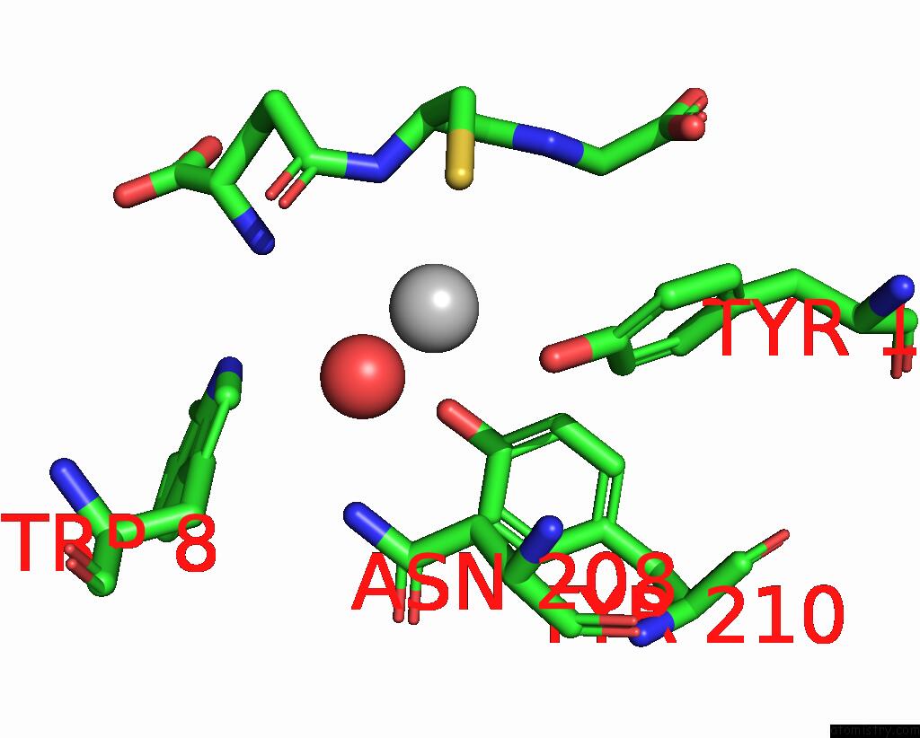
Silver binding site 1 out of 3 in 7so8
Go back to
Silver binding site 1 out
of 3 in the Crystal Structure of Glutathione S-Transferase From Shrimp Litopenaeus Vannamei in Complex with Silver Ions and A Molecules of Glutathione Binding in G-Site and H-Site
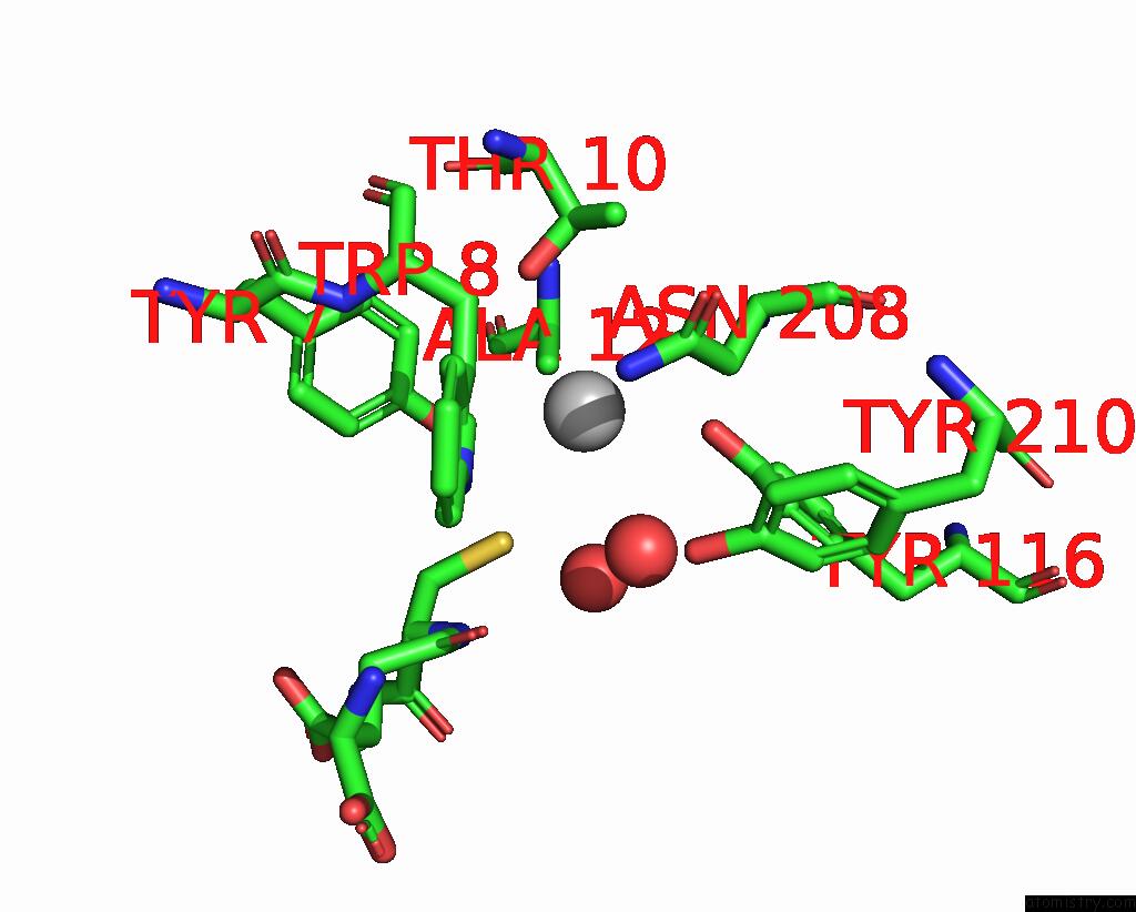
Mono view
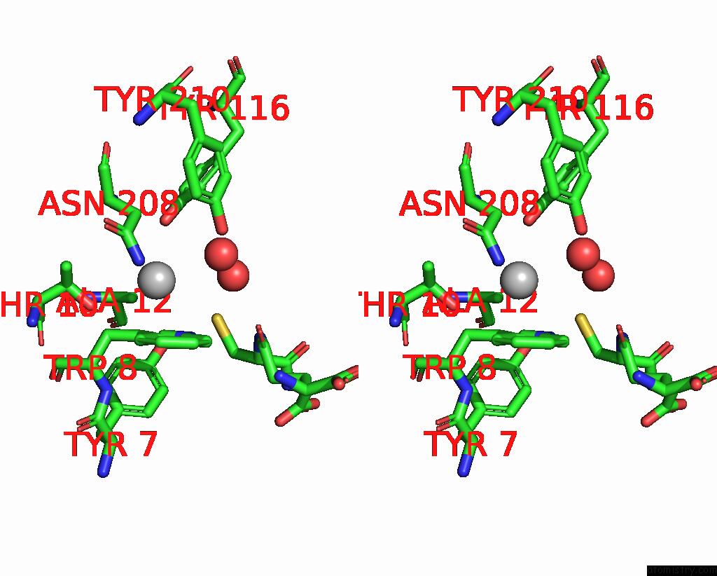
Stereo pair view
Mono view
Stereo pair view
A full contact list of Silver with other atoms in the Ag binding
site number 1 of Crystal Structure of Glutathione S-Transferase From Shrimp Litopenaeus Vannamei in Complex with Silver Ions and A Molecules of Glutathione Binding in G-Site and H-Site within 5.0Å range:
|
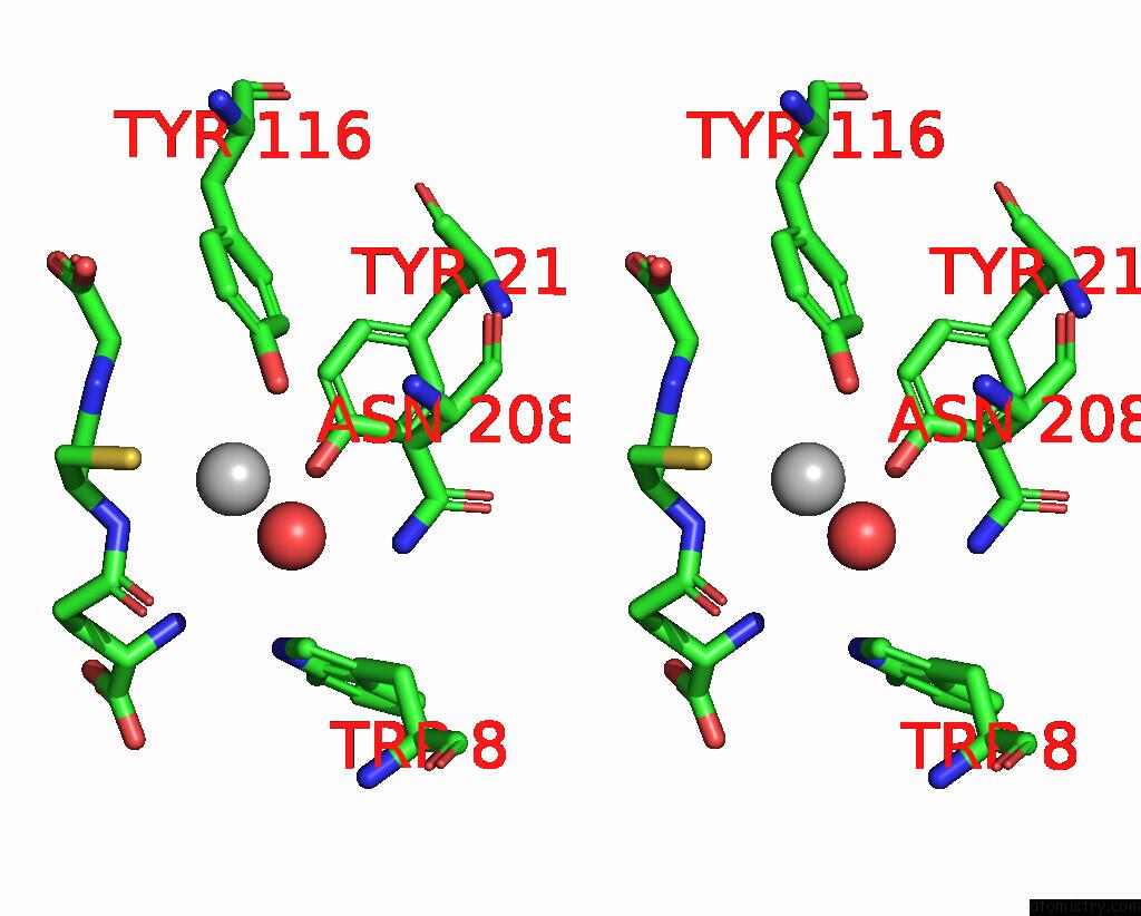
Silver binding site 2 out of 3 in 7so8
Go back to
Silver binding site 2 out
of 3 in the Crystal Structure of Glutathione S-Transferase From Shrimp Litopenaeus Vannamei in Complex with Silver Ions and A Molecules of Glutathione Binding in G-Site and H-Site
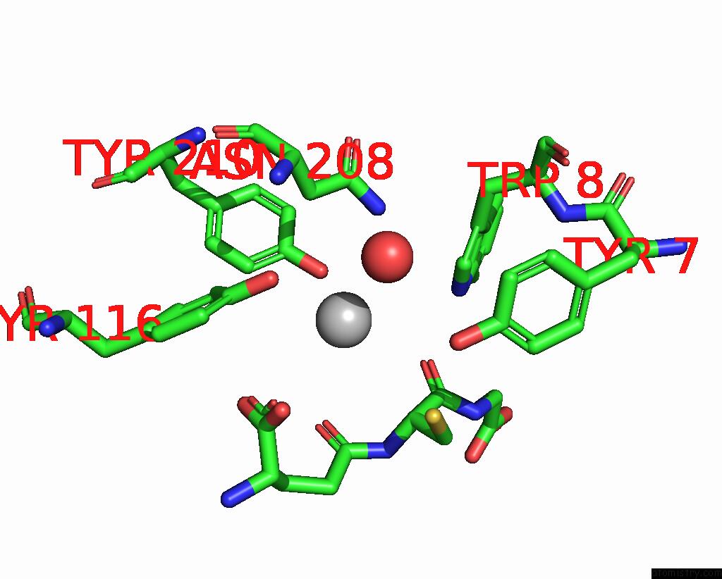
Mono view
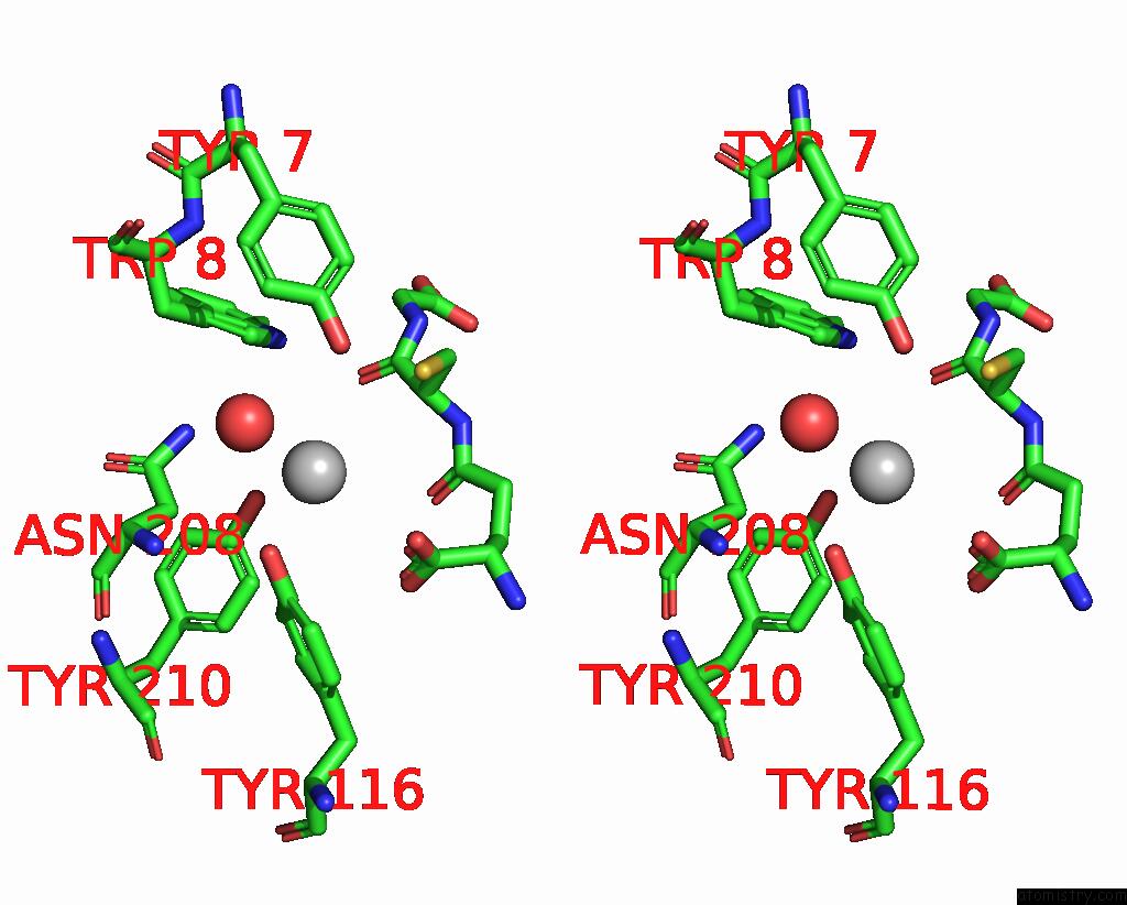
Stereo pair view
Mono view
Stereo pair view
A full contact list of Silver with other atoms in the Ag binding
site number 2 of Crystal Structure of Glutathione S-Transferase From Shrimp Litopenaeus Vannamei in Complex with Silver Ions and A Molecules of Glutathione Binding in G-Site and H-Site within 5.0Å range:
|
Silver binding site 3 out of 3 in 7so8
Go back to
Silver binding site 3 out
of 3 in the Crystal Structure of Glutathione S-Transferase From Shrimp Litopenaeus Vannamei in Complex with Silver Ions and A Molecules of Glutathione Binding in G-Site and H-Site
Mono view
Stereo pair view
Mono view
Stereo pair view
A full contact list of Silver with other atoms in the Ag binding
site number 3 of Crystal Structure of Glutathione S-Transferase From Shrimp Litopenaeus Vannamei in Complex with Silver Ions and A Molecules of Glutathione Binding in G-Site and H-Site within 5.0Å range:
|
Reference:
A.Escudero-Garcia,
E.Rudino-Pinera.
Inhibition of Gst Class Mu of the Shrimp Litopenaeus Vannamei By Binding Silver Ions in H-Site. To Be Published.
Page generated: Sun Jul 6 21:18:10 2025
Last articles
K in 2X21K in 2X6A
K in 2X20
K in 2WOX
K in 2WQY
K in 2WM2
K in 2WLN
K in 2WLK
K in 2WLM
K in 2WLO