Silver »
PDB 1aoo-6ka0 »
2vb3 »
Silver in PDB 2vb3: Crystal Structure of Ag(I)Cusf
Protein crystallography data
The structure of Crystal Structure of Ag(I)Cusf, PDB code: 2vb3
was solved by
Y.Xue,
A.V.Davis,
G.Balakrishnan,
J.P.Stasser,
B.M.Staehlin,
P.Focia,
T.G.Spiro,
J.E.Penner-Hahn,
T.V.O'halloran,
with X-Ray Crystallography technique. A brief refinement statistics is given in the table below:
| Resolution Low / High (Å) | 44.24 / 2.33 |
| Space group | P 21 21 21 |
| Cell size a, b, c (Å), α, β, γ (°) | 39.658, 44.229, 44.241, 90.00, 90.00, 90.00 |
| R / Rfree (%) | 24.1 / 26.4 |
Silver Binding Sites:
The binding sites of Silver atom in the Crystal Structure of Ag(I)Cusf
(pdb code 2vb3). This binding sites where shown within
5.0 Angstroms radius around Silver atom.
In total only one binding site of Silver was determined in the Crystal Structure of Ag(I)Cusf, PDB code: 2vb3:
In total only one binding site of Silver was determined in the Crystal Structure of Ag(I)Cusf, PDB code: 2vb3:
Silver binding site 1 out of 1 in 2vb3
Go back to
Silver binding site 1 out
of 1 in the Crystal Structure of Ag(I)Cusf
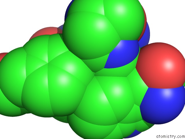
Mono view
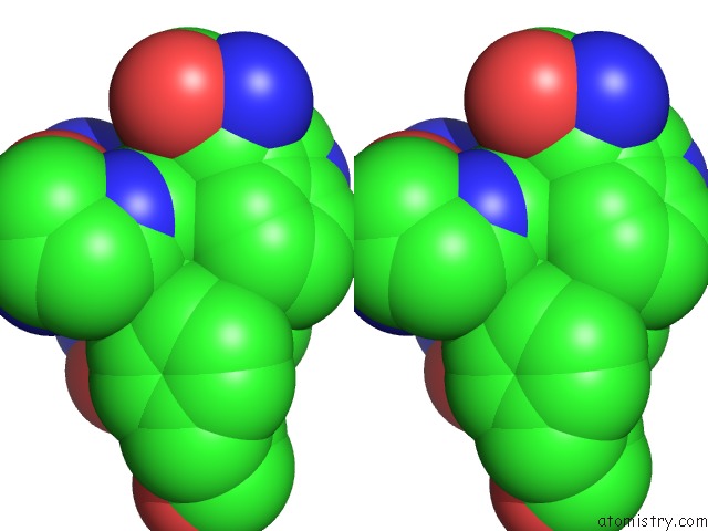
Stereo pair view
Mono view
Stereo pair view
A full contact list of Silver with other atoms in the Ag binding
site number 1 of Crystal Structure of Ag(I)Cusf within 5.0Å range:
|
Reference:
Y.Xue,
A.V.Davis,
G.Balakrishnan,
J.P.Stasser,
B.M.Staehlin,
P.Focia,
T.G.Spiro,
J.E.Penner-Hahn,
T.V.O'halloran.
Cu(I) Recognition Via Cation-Pi and Methionine Interactions in Cusf. Nat.Chem.Biol. V. 4 107 2008.
ISSN: ISSN 1552-4450
PubMed: 18157124
DOI: 10.1038/NCHEMBIO.2007.57
Page generated: Sun Jul 6 20:47:41 2025
ISSN: ISSN 1552-4450
PubMed: 18157124
DOI: 10.1038/NCHEMBIO.2007.57
Last articles
Zn in 4I7CZn in 4I7B
Zn in 4I6V
Zn in 4I68
Zn in 4I5I
Zn in 4I2I
Zn in 4I35
Zn in 4I2X
Zn in 4I2J
Zn in 4I1H