Silver »
PDB 1aoo-6ka0 »
2qcp »
Silver in PDB 2qcp: 1.0 A Structure of Cusf-Ag(I) Residues 10-88 From Escherichia Coli
Protein crystallography data
The structure of 1.0 A Structure of Cusf-Ag(I) Residues 10-88 From Escherichia Coli, PDB code: 2qcp
was solved by
I.R.Loftin,
with X-Ray Crystallography technique. A brief refinement statistics is given in the table below:
| Resolution Low / High (Å) | 19.68 / 1.00 |
| Space group | P 21 21 21 |
| Cell size a, b, c (Å), α, β, γ (°) | 38.118, 39.351, 44.423, 90.00, 90.00, 90.00 |
| R / Rfree (%) | 15.9 / 18.6 |
Silver Binding Sites:
The binding sites of Silver atom in the 1.0 A Structure of Cusf-Ag(I) Residues 10-88 From Escherichia Coli
(pdb code 2qcp). This binding sites where shown within
5.0 Angstroms radius around Silver atom.
In total only one binding site of Silver was determined in the 1.0 A Structure of Cusf-Ag(I) Residues 10-88 From Escherichia Coli, PDB code: 2qcp:
In total only one binding site of Silver was determined in the 1.0 A Structure of Cusf-Ag(I) Residues 10-88 From Escherichia Coli, PDB code: 2qcp:
Silver binding site 1 out of 1 in 2qcp
Go back to
Silver binding site 1 out
of 1 in the 1.0 A Structure of Cusf-Ag(I) Residues 10-88 From Escherichia Coli
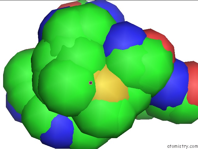
Mono view
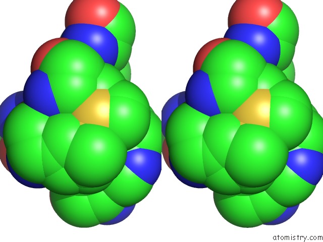
Stereo pair view
Mono view
Stereo pair view
A full contact list of Silver with other atoms in the Ag binding
site number 1 of 1.0 A Structure of Cusf-Ag(I) Residues 10-88 From Escherichia Coli within 5.0Å range:
|
Reference:
I.R.Loftin,
S.Franke,
N.J.Blackburn,
M.M.Mcevoy.
Unusual Cu(I)/Ag(I) Coordination of Escherichia Coli Cusf As Revealed By Atomic Resolution Crystallography and X-Ray Absorption Spectroscopy Protein Sci. V. 16 2287 2007.
ISSN: ISSN 0961-8368
PubMed: 17893365
DOI: 10.1110/PS.073021307
Page generated: Sun Jul 6 20:47:32 2025
ISSN: ISSN 0961-8368
PubMed: 17893365
DOI: 10.1110/PS.073021307
Last articles
Zn in 4ICQZn in 4IDR
Zn in 4IC3
Zn in 4IC2
Zn in 4IBZ
Zn in 4IBY
Zn in 4IBW
Zn in 4IBV
Zn in 4IBU
Zn in 4IBQ