Silver »
PDB 7sdv-8dx7 »
7so8 »
Silver in PDB 7so8: Crystal Structure of Glutathione S-Transferase From Shrimp Litopenaeus Vannamei in Complex with Silver Ions and A Molecules of Glutathione Binding in G-Site and H-Site
Enzymatic activity of Crystal Structure of Glutathione S-Transferase From Shrimp Litopenaeus Vannamei in Complex with Silver Ions and A Molecules of Glutathione Binding in G-Site and H-Site
All present enzymatic activity of Crystal Structure of Glutathione S-Transferase From Shrimp Litopenaeus Vannamei in Complex with Silver Ions and A Molecules of Glutathione Binding in G-Site and H-Site:
2.5.1.18;
2.5.1.18;
Protein crystallography data
The structure of Crystal Structure of Glutathione S-Transferase From Shrimp Litopenaeus Vannamei in Complex with Silver Ions and A Molecules of Glutathione Binding in G-Site and H-Site, PDB code: 7so8
was solved by
A.Escudero-Garcia,
E.Rudino-Pinera,
R.Miranda-Blancas,
with X-Ray Crystallography technique. A brief refinement statistics is given in the table below:
| Resolution Low / High (Å) | 56.37 / 2.20 |
| Space group | P 1 21 1 |
| Cell size a, b, c (Å), α, β, γ (°) | 57.39, 93.02, 169.11, 90, 90.75, 90 |
| R / Rfree (%) | 19.1 / 26 |
Silver Binding Sites:
The binding sites of Silver atom in the Crystal Structure of Glutathione S-Transferase From Shrimp Litopenaeus Vannamei in Complex with Silver Ions and A Molecules of Glutathione Binding in G-Site and H-Site
(pdb code 7so8). This binding sites where shown within
5.0 Angstroms radius around Silver atom.
In total 3 binding sites of Silver where determined in the Crystal Structure of Glutathione S-Transferase From Shrimp Litopenaeus Vannamei in Complex with Silver Ions and A Molecules of Glutathione Binding in G-Site and H-Site, PDB code: 7so8:
Jump to Silver binding site number: 1; 2; 3;
In total 3 binding sites of Silver where determined in the Crystal Structure of Glutathione S-Transferase From Shrimp Litopenaeus Vannamei in Complex with Silver Ions and A Molecules of Glutathione Binding in G-Site and H-Site, PDB code: 7so8:
Jump to Silver binding site number: 1; 2; 3;
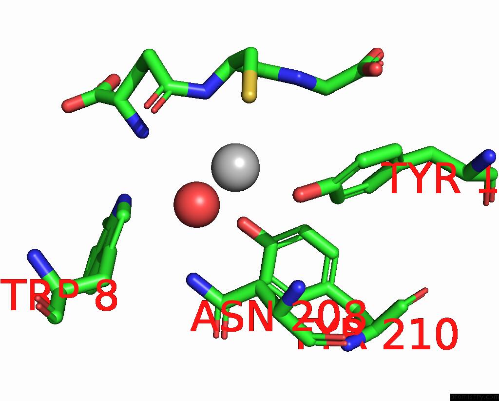
Silver binding site 1 out of 3 in 7so8
Go back to
Silver binding site 1 out
of 3 in the Crystal Structure of Glutathione S-Transferase From Shrimp Litopenaeus Vannamei in Complex with Silver Ions and A Molecules of Glutathione Binding in G-Site and H-Site
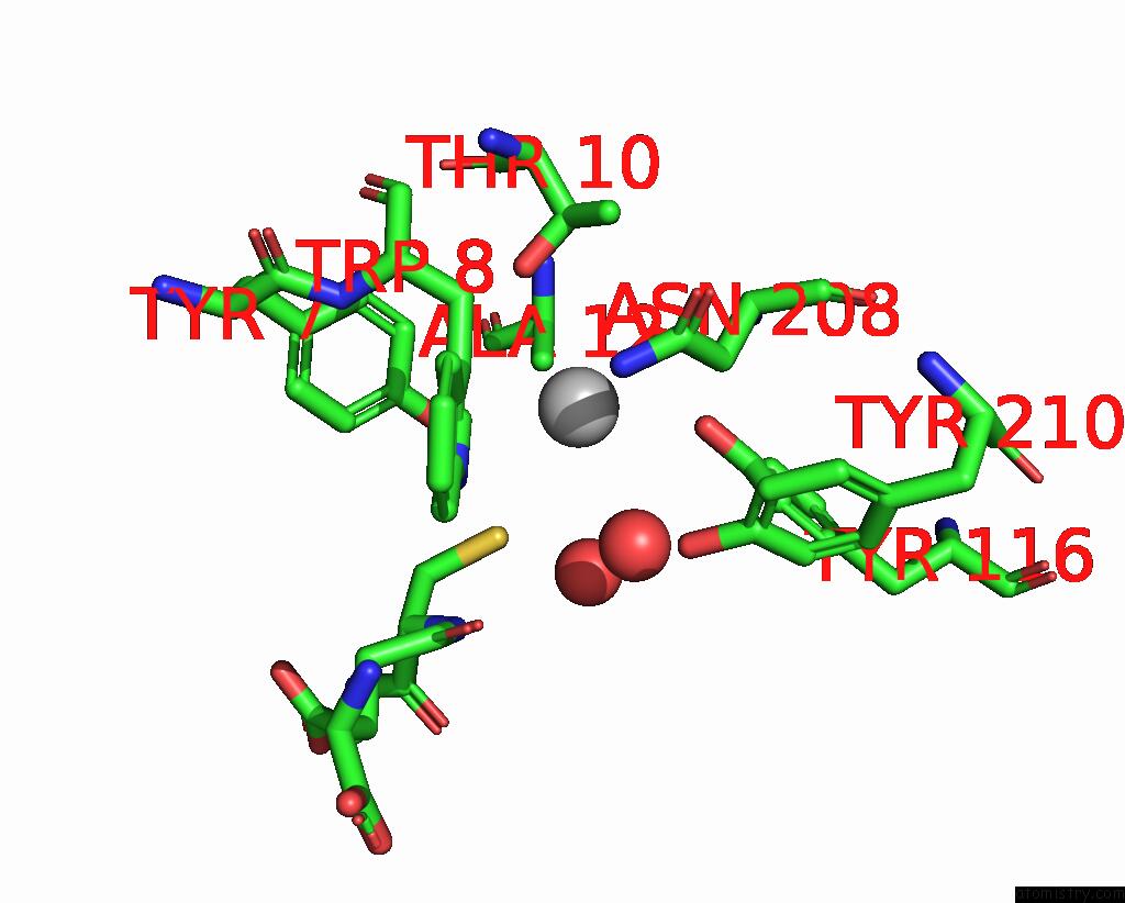
Mono view
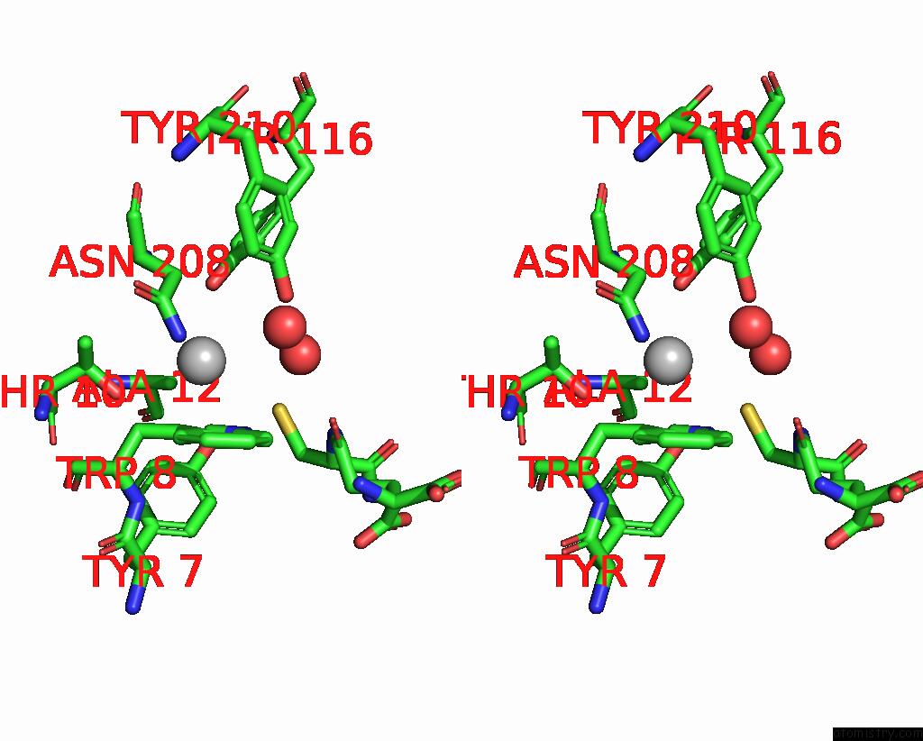
Stereo pair view
Mono view
Stereo pair view
A full contact list of Silver with other atoms in the Ag binding
site number 1 of Crystal Structure of Glutathione S-Transferase From Shrimp Litopenaeus Vannamei in Complex with Silver Ions and A Molecules of Glutathione Binding in G-Site and H-Site within 5.0Å range:
|
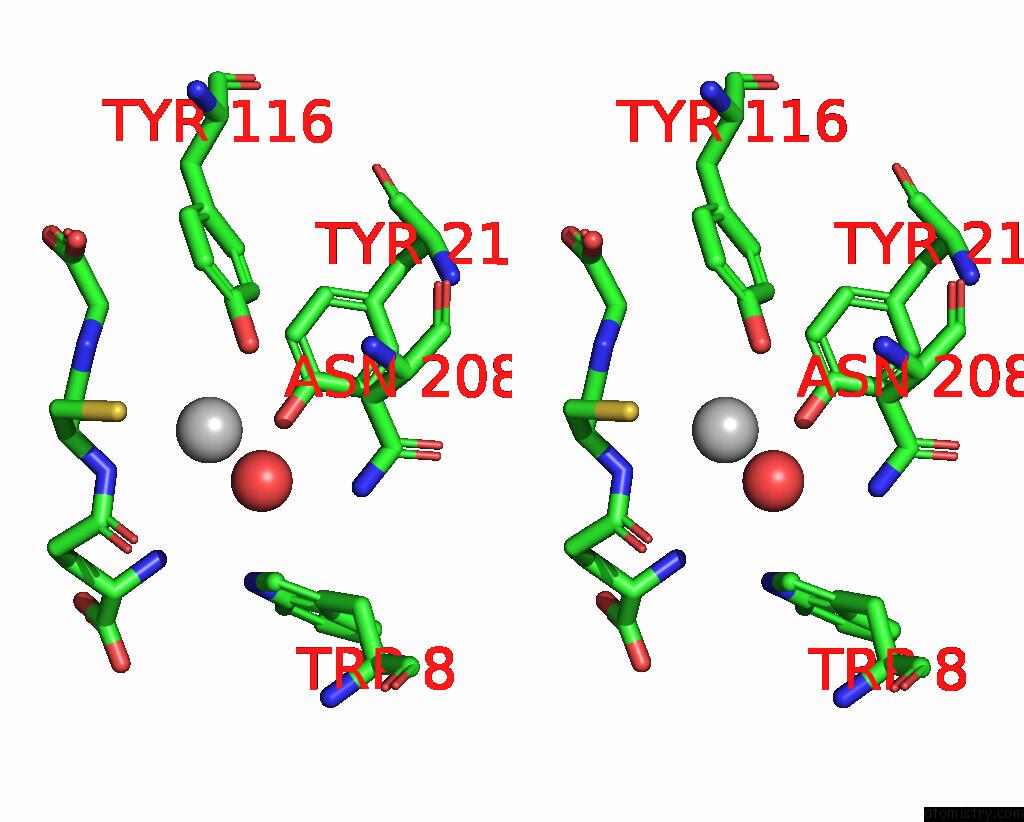
Silver binding site 2 out of 3 in 7so8
Go back to
Silver binding site 2 out
of 3 in the Crystal Structure of Glutathione S-Transferase From Shrimp Litopenaeus Vannamei in Complex with Silver Ions and A Molecules of Glutathione Binding in G-Site and H-Site
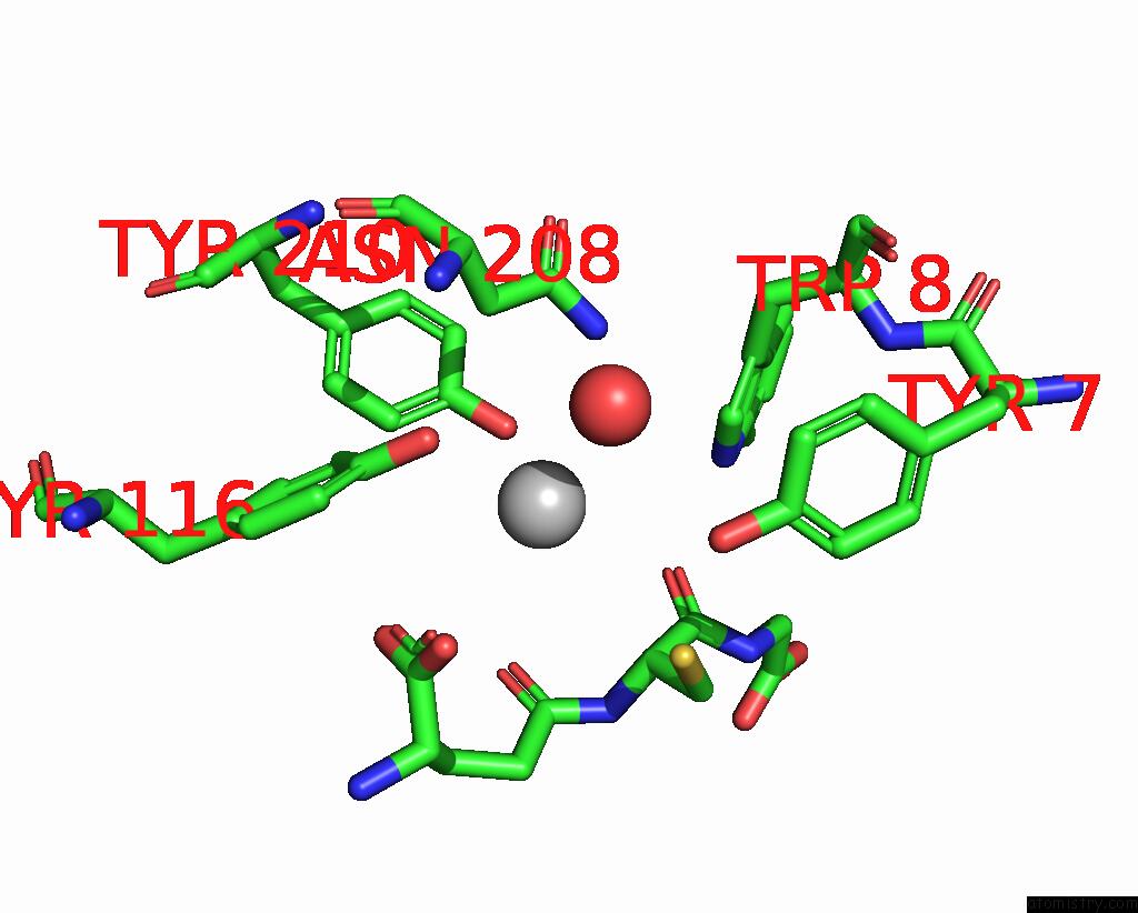
Mono view
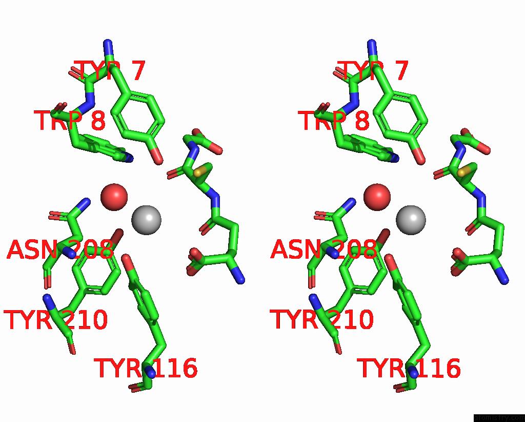
Stereo pair view
Mono view
Stereo pair view
A full contact list of Silver with other atoms in the Ag binding
site number 2 of Crystal Structure of Glutathione S-Transferase From Shrimp Litopenaeus Vannamei in Complex with Silver Ions and A Molecules of Glutathione Binding in G-Site and H-Site within 5.0Å range:
|
Silver binding site 3 out of 3 in 7so8
Go back to
Silver binding site 3 out
of 3 in the Crystal Structure of Glutathione S-Transferase From Shrimp Litopenaeus Vannamei in Complex with Silver Ions and A Molecules of Glutathione Binding in G-Site and H-Site
Mono view
Stereo pair view
Mono view
Stereo pair view
A full contact list of Silver with other atoms in the Ag binding
site number 3 of Crystal Structure of Glutathione S-Transferase From Shrimp Litopenaeus Vannamei in Complex with Silver Ions and A Molecules of Glutathione Binding in G-Site and H-Site within 5.0Å range:
|
Reference:
A.Escudero-Garcia,
E.Rudino-Pinera.
Inhibition of Gst Class Mu of the Shrimp Litopenaeus Vannamei By Binding Silver Ions in H-Site. To Be Published.
Page generated: Wed Jul 10 09:02:05 2024
Last articles
Zn in 9MJ5Zn in 9HNW
Zn in 9G0L
Zn in 9FNE
Zn in 9DZN
Zn in 9E0I
Zn in 9D32
Zn in 9DAK
Zn in 8ZXC
Zn in 8ZUF