Silver »
PDB 1aoo-6ka0 »
5xuv »
Silver in PDB 5xuv: Crystal Structure of Dna Duplex Containing 4-Thiothymine-2AG(I)-4- Thiothymine Base Pairs
Protein crystallography data
The structure of Crystal Structure of Dna Duplex Containing 4-Thiothymine-2AG(I)-4- Thiothymine Base Pairs, PDB code: 5xuv
was solved by
J.Kondo,
T.Sugawara,
H.Saneyoshi,
A.Ono,
with X-Ray Crystallography technique. A brief refinement statistics is given in the table below:
| Resolution Low / High (Å) | 33.22 / 1.90 |
| Space group | P 21 21 21 |
| Cell size a, b, c (Å), α, β, γ (°) | 25.213, 40.487, 66.432, 90.00, 90.00, 90.00 |
| R / Rfree (%) | 18.5 / 22.2 |
Other elements in 5xuv:
The structure of Crystal Structure of Dna Duplex Containing 4-Thiothymine-2AG(I)-4- Thiothymine Base Pairs also contains other interesting chemical elements:
| Potassium | (K) | 2 atoms |
Silver Binding Sites:
The binding sites of Silver atom in the Crystal Structure of Dna Duplex Containing 4-Thiothymine-2AG(I)-4- Thiothymine Base Pairs
(pdb code 5xuv). This binding sites where shown within
5.0 Angstroms radius around Silver atom.
In total 4 binding sites of Silver where determined in the Crystal Structure of Dna Duplex Containing 4-Thiothymine-2AG(I)-4- Thiothymine Base Pairs, PDB code: 5xuv:
Jump to Silver binding site number: 1; 2; 3; 4;
In total 4 binding sites of Silver where determined in the Crystal Structure of Dna Duplex Containing 4-Thiothymine-2AG(I)-4- Thiothymine Base Pairs, PDB code: 5xuv:
Jump to Silver binding site number: 1; 2; 3; 4;
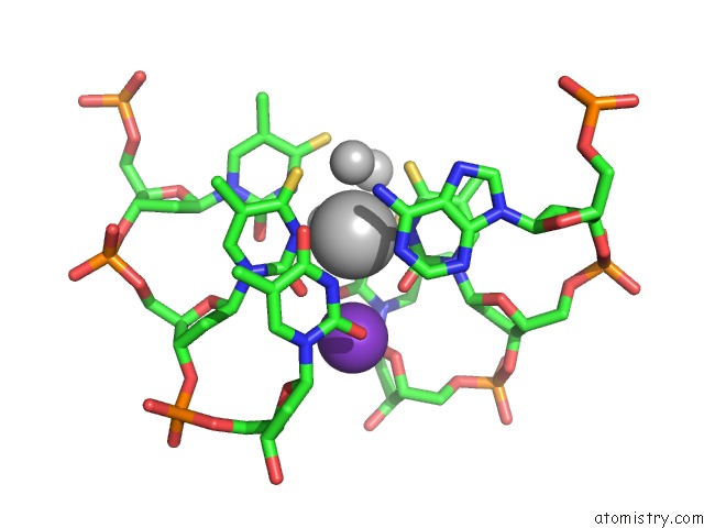
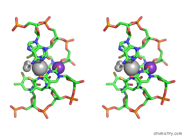
Silver binding site 1 out of 4 in 5xuv
Go back to
Silver binding site 1 out
of 4 in the Crystal Structure of Dna Duplex Containing 4-Thiothymine-2AG(I)-4- Thiothymine Base Pairs
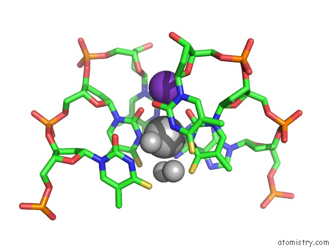
Mono view
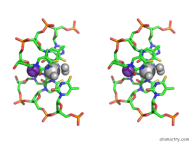
Stereo pair view
Mono view
Stereo pair view
A full contact list of Silver with other atoms in the Ag binding
site number 1 of Crystal Structure of Dna Duplex Containing 4-Thiothymine-2AG(I)-4- Thiothymine Base Pairs within 5.0Å range:
|
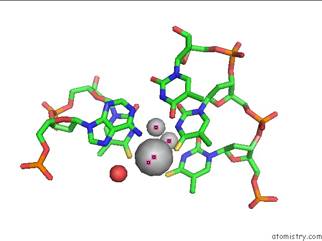
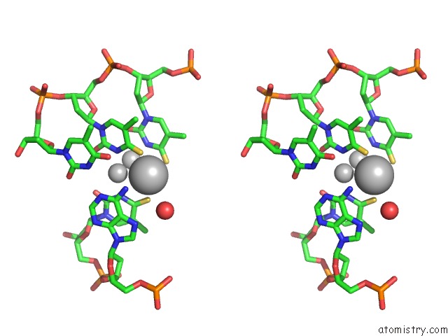
Silver binding site 2 out of 4 in 5xuv
Go back to
Silver binding site 2 out
of 4 in the Crystal Structure of Dna Duplex Containing 4-Thiothymine-2AG(I)-4- Thiothymine Base Pairs
Mono view
Stereo pair view
Mono view
Stereo pair view
A full contact list of Silver with other atoms in the Ag binding
site number 2 of Crystal Structure of Dna Duplex Containing 4-Thiothymine-2AG(I)-4- Thiothymine Base Pairs within 5.0Å range:
|
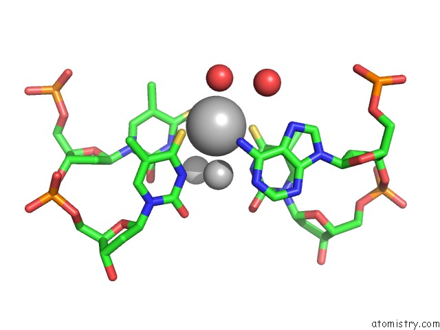
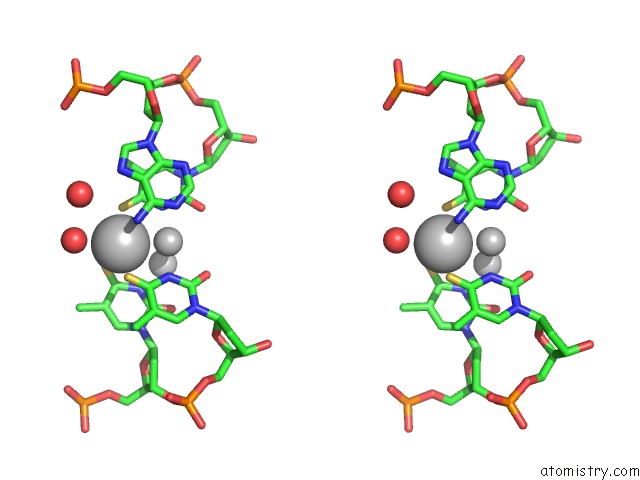
Silver binding site 3 out of 4 in 5xuv
Go back to
Silver binding site 3 out
of 4 in the Crystal Structure of Dna Duplex Containing 4-Thiothymine-2AG(I)-4- Thiothymine Base Pairs
Mono view
Stereo pair view
Mono view
Stereo pair view
A full contact list of Silver with other atoms in the Ag binding
site number 3 of Crystal Structure of Dna Duplex Containing 4-Thiothymine-2AG(I)-4- Thiothymine Base Pairs within 5.0Å range:
|
Silver binding site 4 out of 4 in 5xuv
Go back to
Silver binding site 4 out
of 4 in the Crystal Structure of Dna Duplex Containing 4-Thiothymine-2AG(I)-4- Thiothymine Base Pairs
Mono view
Stereo pair view
Mono view
Stereo pair view
A full contact list of Silver with other atoms in the Ag binding
site number 4 of Crystal Structure of Dna Duplex Containing 4-Thiothymine-2AG(I)-4- Thiothymine Base Pairs within 5.0Å range:
|
Reference:
J.Kondo,
T.Sugawara,
H.Saneyoshi,
A.Ono.
Crystal Structure of A Dna Duplex Containing Four Ag(I) Ions in Consecutive Dinuclear Ag(I)-Mediated Base Pairs: 4-Thiothymine-2AG(I)-4-Thiothymine Chem. Commun. (Camb.) V. 53 11747 2017.
ISSN: ESSN 1364-548X
PubMed: 28991300
DOI: 10.1039/C7CC06153F
Page generated: Wed Jul 10 08:26:40 2024
ISSN: ESSN 1364-548X
PubMed: 28991300
DOI: 10.1039/C7CC06153F
Last articles
Zn in 9J0NZn in 9J0O
Zn in 9J0P
Zn in 9FJX
Zn in 9EKB
Zn in 9C0F
Zn in 9CAH
Zn in 9CH0
Zn in 9CH3
Zn in 9CH1