Silver »
PDB 1aoo-6ka0 »
2x17 »
Silver in PDB 2x17: The X-Ray Structure of Ferritin From Pyrococcus Furiosus Loaded with Ag(I)
Protein crystallography data
The structure of The X-Ray Structure of Ferritin From Pyrococcus Furiosus Loaded with Ag(I), PDB code: 2x17
was solved by
A.Ilari,
A.Fiorillo,
P.Ceci,
with X-Ray Crystallography technique. A brief refinement statistics is given in the table below:
| Resolution Low / High (Å) | 50.00 / 3.10 |
| Space group | P 41 |
| Cell size a, b, c (Å), α, β, γ (°) | 157.681, 157.681, 246.437, 90.00, 90.00, 90.00 |
| R / Rfree (%) | 26.014 / 28.338 |
Silver Binding Sites:
Pages:
>>> Page 1 <<< Page 2, Binding sites: 11 - 20; Page 3, Binding sites: 21 - 30; Page 4, Binding sites: 31 - 33;Binding sites:
The binding sites of Silver atom in the The X-Ray Structure of Ferritin From Pyrococcus Furiosus Loaded with Ag(I) (pdb code 2x17). This binding sites where shown within 5.0 Angstroms radius around Silver atom.In total 33 binding sites of Silver where determined in the The X-Ray Structure of Ferritin From Pyrococcus Furiosus Loaded with Ag(I), PDB code: 2x17:
Jump to Silver binding site number: 1; 2; 3; 4; 5; 6; 7; 8; 9; 10;
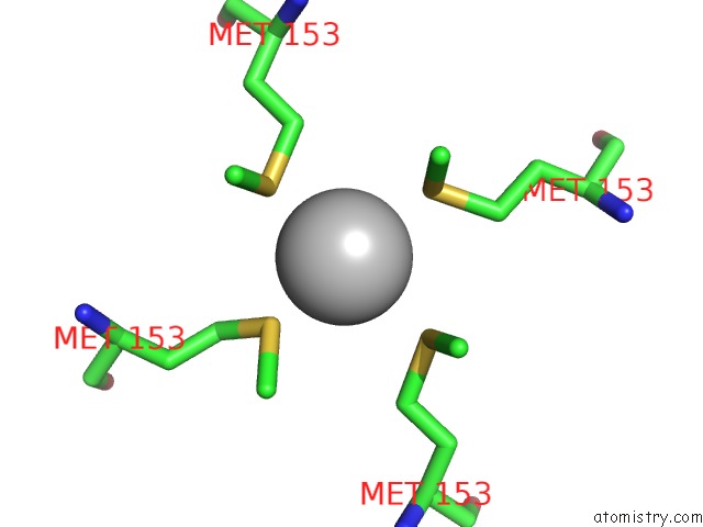
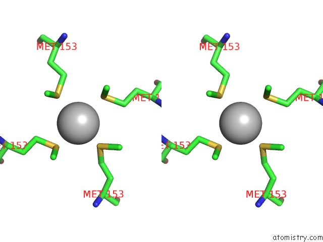
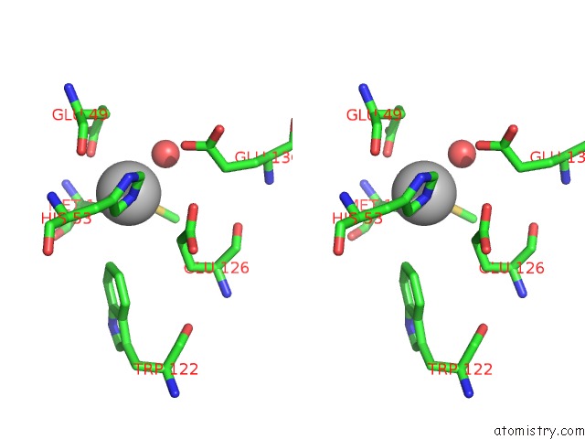
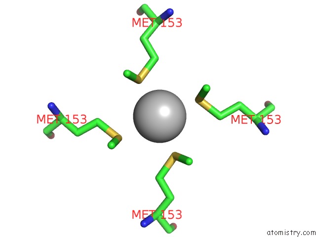
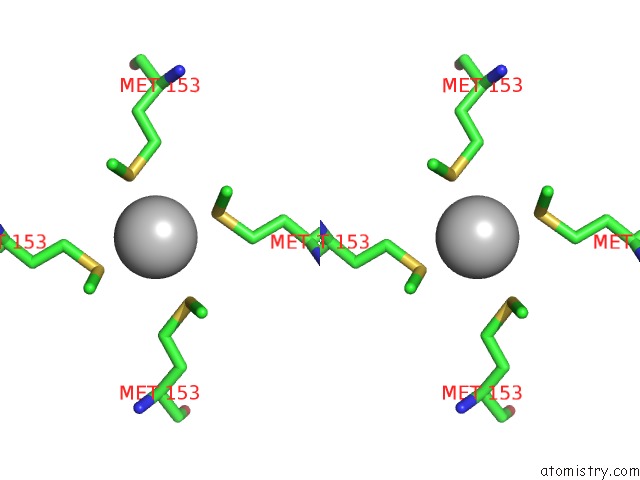
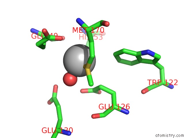
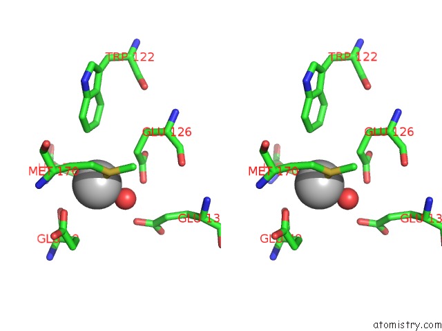
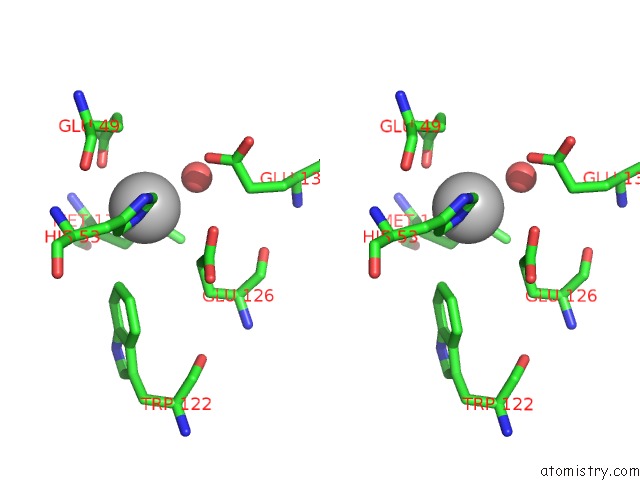
Silver binding site 1 out of 33 in 2x17
Go back to
Silver binding site 1 out
of 33 in the The X-Ray Structure of Ferritin From Pyrococcus Furiosus Loaded with Ag(I)
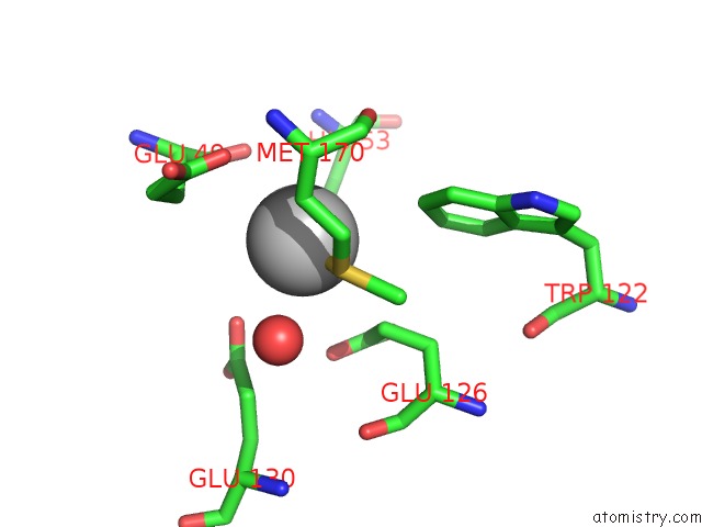
Mono view
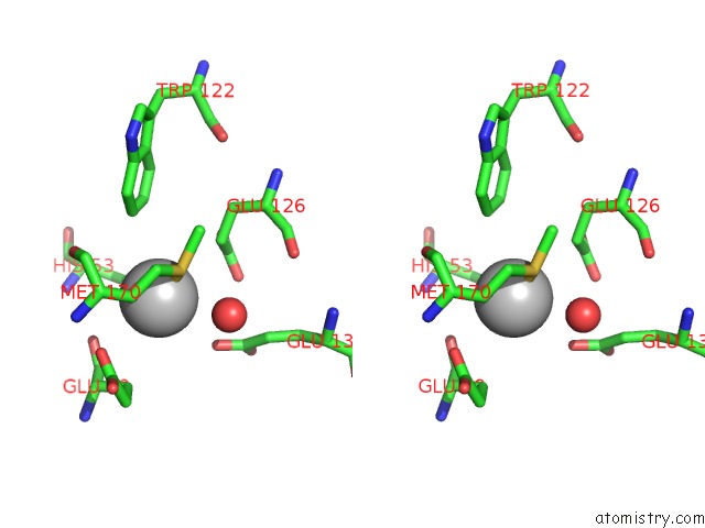
Stereo pair view
Mono view
Stereo pair view
A full contact list of Silver with other atoms in the Ag binding
site number 1 of The X-Ray Structure of Ferritin From Pyrococcus Furiosus Loaded with Ag(I) within 5.0Å range:
|
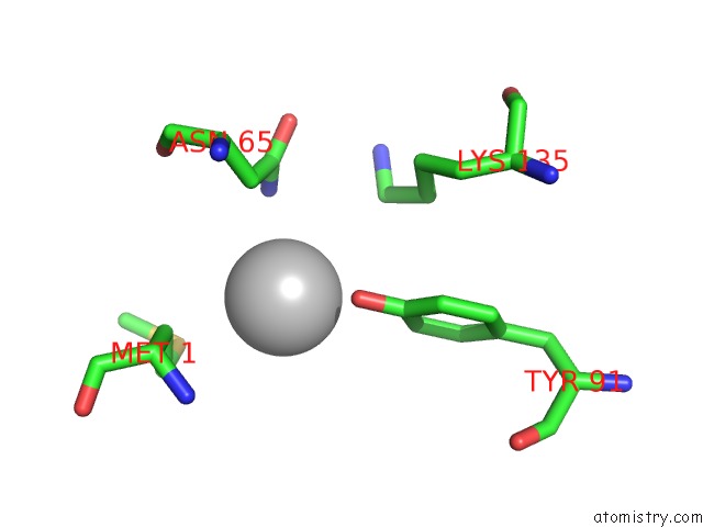
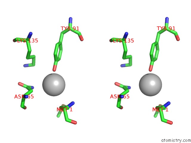
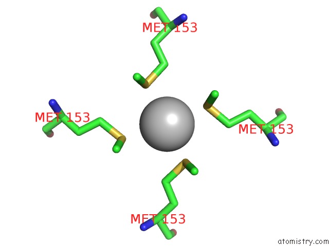
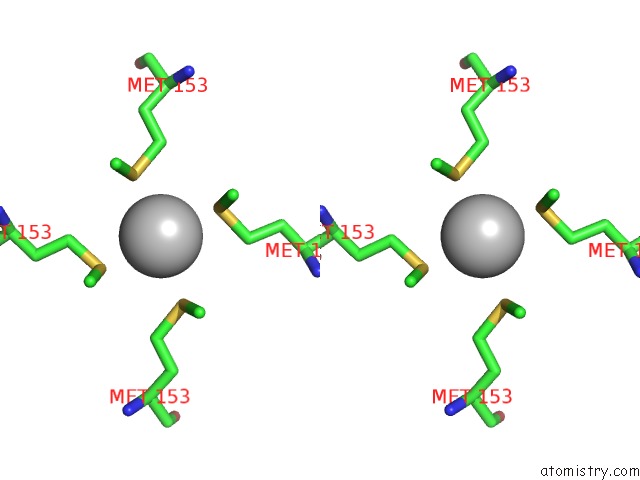
Silver binding site 2 out of 33 in 2x17
Go back to
Silver binding site 2 out
of 33 in the The X-Ray Structure of Ferritin From Pyrococcus Furiosus Loaded with Ag(I)
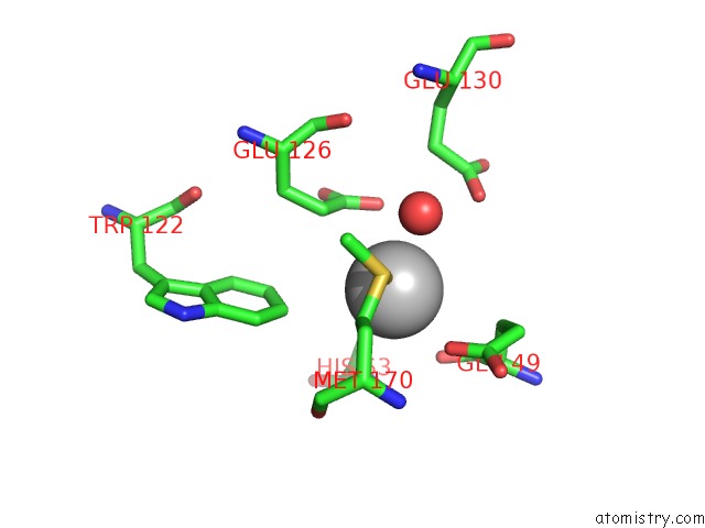
Mono view
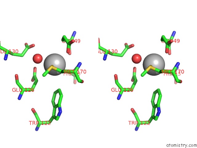
Stereo pair view
Mono view
Stereo pair view
A full contact list of Silver with other atoms in the Ag binding
site number 2 of The X-Ray Structure of Ferritin From Pyrococcus Furiosus Loaded with Ag(I) within 5.0Å range:
|
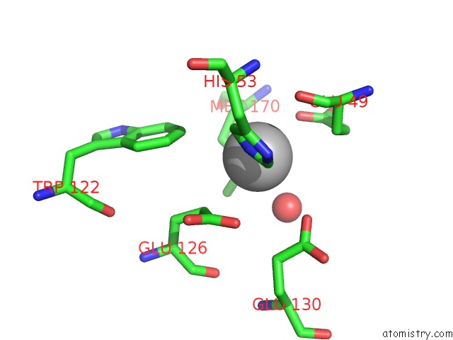
Silver binding site 3 out of 33 in 2x17
Go back to
Silver binding site 3 out
of 33 in the The X-Ray Structure of Ferritin From Pyrococcus Furiosus Loaded with Ag(I)
Mono view
Stereo pair view
Mono view
Stereo pair view
A full contact list of Silver with other atoms in the Ag binding
site number 3 of The X-Ray Structure of Ferritin From Pyrococcus Furiosus Loaded with Ag(I) within 5.0Å range:
|
Silver binding site 4 out of 33 in 2x17
Go back to
Silver binding site 4 out
of 33 in the The X-Ray Structure of Ferritin From Pyrococcus Furiosus Loaded with Ag(I)
Mono view

Stereo pair view
Mono view
Stereo pair view
A full contact list of Silver with other atoms in the Ag binding
site number 4 of The X-Ray Structure of Ferritin From Pyrococcus Furiosus Loaded with Ag(I) within 5.0Å range:
|
Silver binding site 5 out of 33 in 2x17
Go back to
Silver binding site 5 out
of 33 in the The X-Ray Structure of Ferritin From Pyrococcus Furiosus Loaded with Ag(I)
Mono view
Stereo pair view
Mono view
Stereo pair view
A full contact list of Silver with other atoms in the Ag binding
site number 5 of The X-Ray Structure of Ferritin From Pyrococcus Furiosus Loaded with Ag(I) within 5.0Å range:
|
Silver binding site 6 out of 33 in 2x17
Go back to
Silver binding site 6 out
of 33 in the The X-Ray Structure of Ferritin From Pyrococcus Furiosus Loaded with Ag(I)
Mono view
Stereo pair view
Mono view
Stereo pair view
A full contact list of Silver with other atoms in the Ag binding
site number 6 of The X-Ray Structure of Ferritin From Pyrococcus Furiosus Loaded with Ag(I) within 5.0Å range:
|
Silver binding site 7 out of 33 in 2x17
Go back to
Silver binding site 7 out
of 33 in the The X-Ray Structure of Ferritin From Pyrococcus Furiosus Loaded with Ag(I)
Mono view
Stereo pair view
Mono view
Stereo pair view
A full contact list of Silver with other atoms in the Ag binding
site number 7 of The X-Ray Structure of Ferritin From Pyrococcus Furiosus Loaded with Ag(I) within 5.0Å range:
|
Silver binding site 8 out of 33 in 2x17
Go back to
Silver binding site 8 out
of 33 in the The X-Ray Structure of Ferritin From Pyrococcus Furiosus Loaded with Ag(I)
Mono view
Stereo pair view
Mono view
Stereo pair view
A full contact list of Silver with other atoms in the Ag binding
site number 8 of The X-Ray Structure of Ferritin From Pyrococcus Furiosus Loaded with Ag(I) within 5.0Å range:
|
Silver binding site 9 out of 33 in 2x17
Go back to
Silver binding site 9 out
of 33 in the The X-Ray Structure of Ferritin From Pyrococcus Furiosus Loaded with Ag(I)
Mono view
Stereo pair view
Mono view
Stereo pair view
A full contact list of Silver with other atoms in the Ag binding
site number 9 of The X-Ray Structure of Ferritin From Pyrococcus Furiosus Loaded with Ag(I) within 5.0Å range:
|
Silver binding site 10 out of 33 in 2x17
Go back to
Silver binding site 10 out
of 33 in the The X-Ray Structure of Ferritin From Pyrococcus Furiosus Loaded with Ag(I)
Mono view
Stereo pair view
Mono view
Stereo pair view
A full contact list of Silver with other atoms in the Ag binding
site number 10 of The X-Ray Structure of Ferritin From Pyrococcus Furiosus Loaded with Ag(I) within 5.0Å range:
|
Reference:
O.Kasyutich,
A.Ilari,
A.Fiorillo,
D.Tatchev,
A.Hoell,
P.Ceci.
Silver Ion Incorporation and Nanoparticle Formation Inside the Cavity of Pyrococcus Furiosus Ferritin: Structural and Size-Distribution Analyses. J.Am.Chem.Soc. V. 132 3621 2010.
ISSN: ISSN 0002-7863
PubMed: 20170158
DOI: 10.1021/JA910918B
Page generated: Wed Jul 10 08:21:14 2024
ISSN: ISSN 0002-7863
PubMed: 20170158
DOI: 10.1021/JA910918B
Last articles
Zn in 9J0NZn in 9J0O
Zn in 9J0P
Zn in 9FJX
Zn in 9EKB
Zn in 9C0F
Zn in 9CAH
Zn in 9CH0
Zn in 9CH3
Zn in 9CH1